Audition and Somatosensation Anatomy and Physiology I
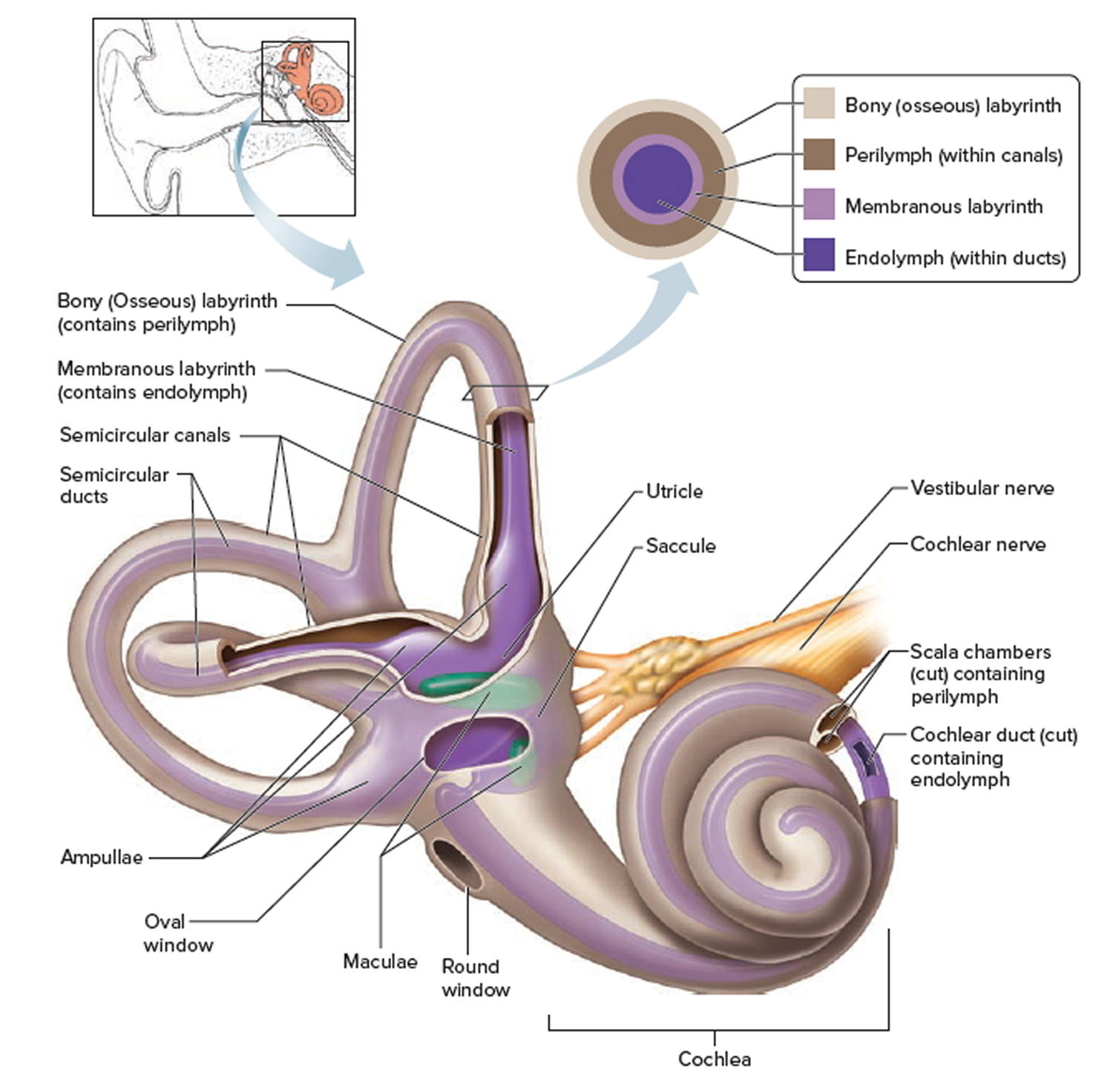
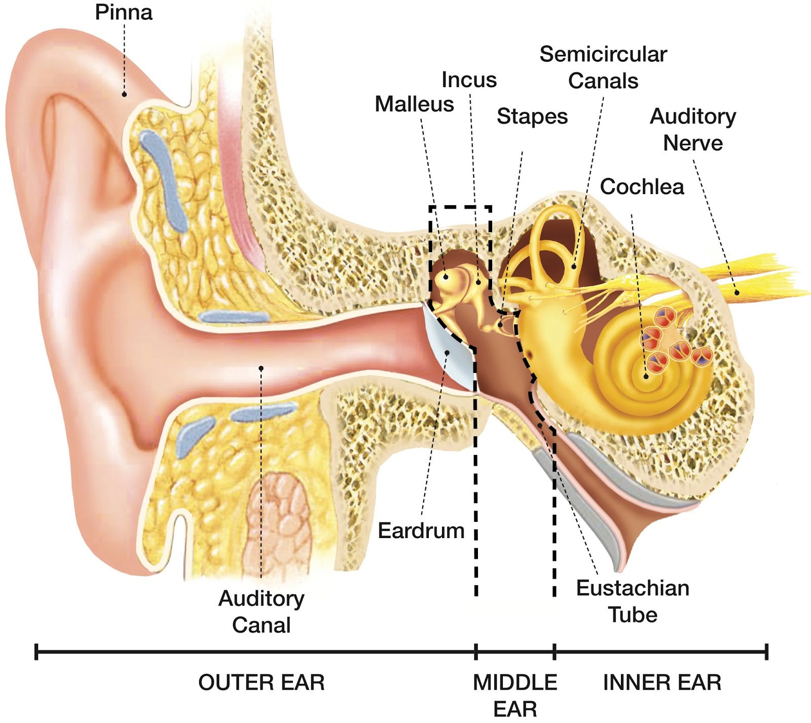
The purpose of the inner ear is to sense and process information about sound and balance, and send that information to the brain. Each part of the inner ear has a specific function. Cochlea: The cochlea is responsible for hearing. It is made up of several layers, with the Organ of Corti at the center.
Inner Ear Problems Causes & Treatment of inner ear Dizziness & Vertigo
The inner ear is the innermost part of the ear and consists of the cochlea, auditory nerve, vestibule and semicircular canals. The inner ear is a maze of tubes and passages, referred to as the labyrinth. The inner ear is mainly responsible for balance and detecting sound. The cochlea contains the cells responsible for hearing, the auditory.
30 Ear Diagram With Label Labels Design Ideas 2020
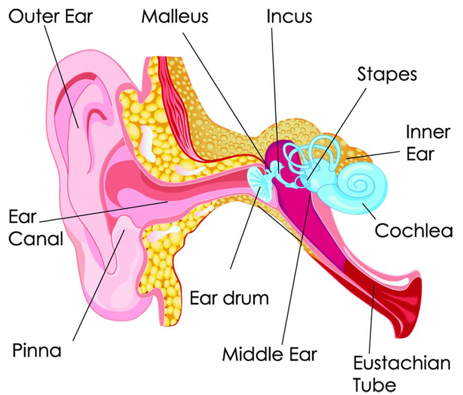
The Structure of Human Ear. Helix: It is the prominent outer rim of the external ear. Antihelix: It is the cartilage curve that is situated parallel to the helix. Crus of the Helix: It is the landmark of the outer ear, situated right above the pointy protrusion known as the tragus. Auditory Ossicles: The three small bones in the middle ear.

Disorders of the Ear Part Two a PA Review and Podcast
The external ear is the visible part of the hearing apparatus. It is comprised of the auricle (pinna) and external auditory canal, including the lateral surface of the tympanic membrane. Together with the tympanic membrane and the middle ear, the pinna serves to amplify sound. The pinna acts as a funnel to deliver sound to the external acoustic meatus, and the external auditory canal.
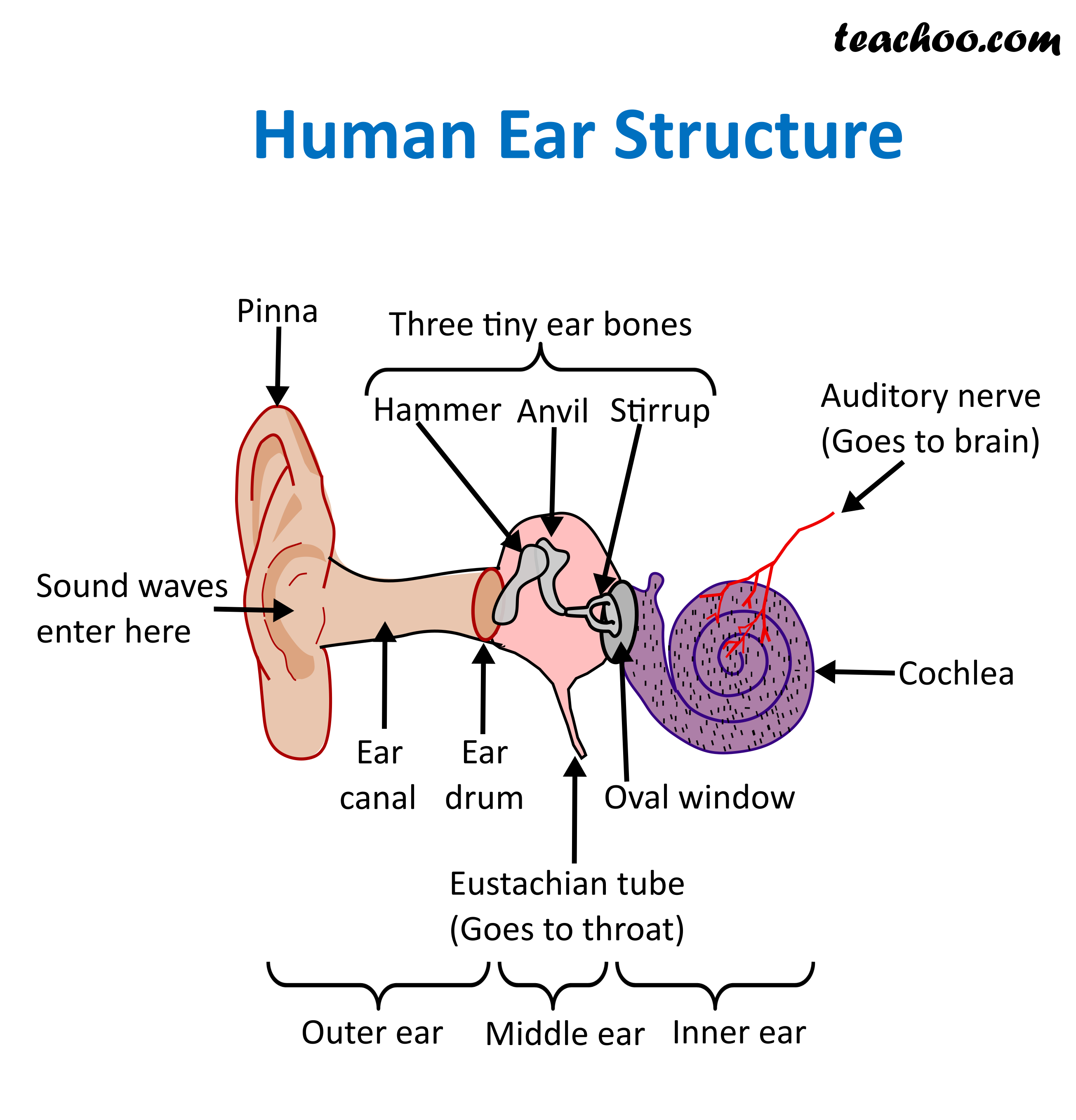
Structure and Function of Human Ear with Diagram Teachoo
Your inner ear contains two main parts: the cochlea and the semicircular canals. Your cochlea is the hearing organ. This snail-shaped structure contains two fluid-filled chambers lined with tiny hairs. When sound enters, the fluid inside of your cochlea causes the tiny hairs to vibrate, sending electrical impulses to your brain.

HEARING ANATOMY AND PROCESS AUDIOLOGIS
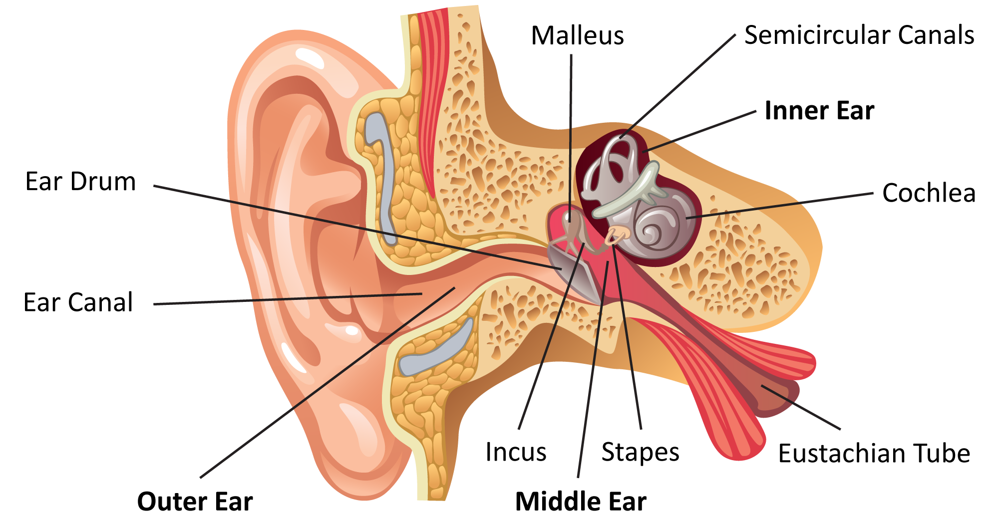
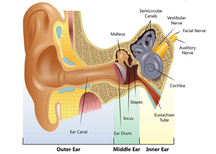
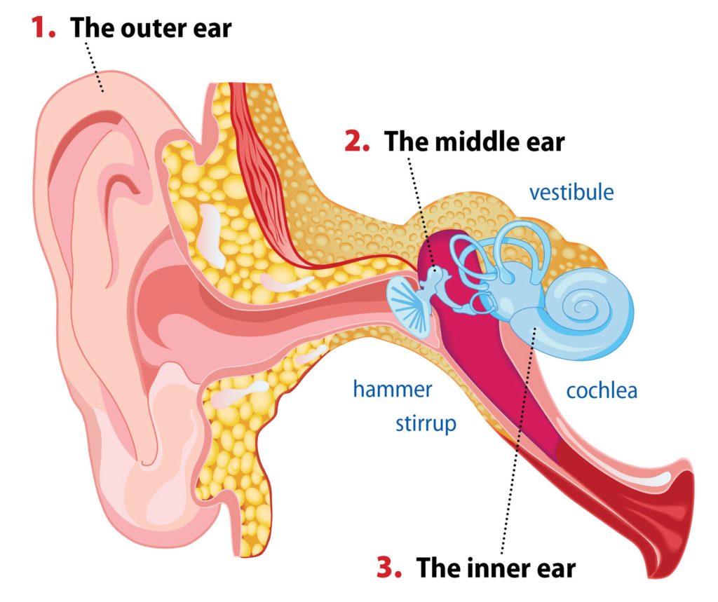
The ear is a complex part of an even more complex sensory system. It is situated bilaterally on the human skull, at the same level as the nose. The main functions of the ear are, of course, hearing, as well as constantly maintaining balance. The ear is anatomically divided into three portions: External ear. Middle ear.
How We Perceive Sound
The ear canal connects the outer cartilage of the ear to the eardrum, which allows people to hear. Read on to learn more about the ear canal.. Anatomy, head and neck, ear. https://www.ncbi.nlm.

Hearing Loss Regenerated in Damaged Mammal Ear The Personal Longevity
Ear anatomy can vary. In addition to normal and relatively minor differences, there are a number of more significant and impactful variants. For instance, on the auricle, attachment—or lack thereof—of the earlobe to the face is a frequently seen genetic variation, with attached earlobes seen in anywhere from 19% to 54% of the population..

Vertigo Have You Spinning Chiropractic Home Care Ear anatomy, Human
A bony casing houses a complex system of membranous cells. The inner ear is called the labyrinth because of its complex shape. There are two main sections within the inner ear: the bony labyrinth.
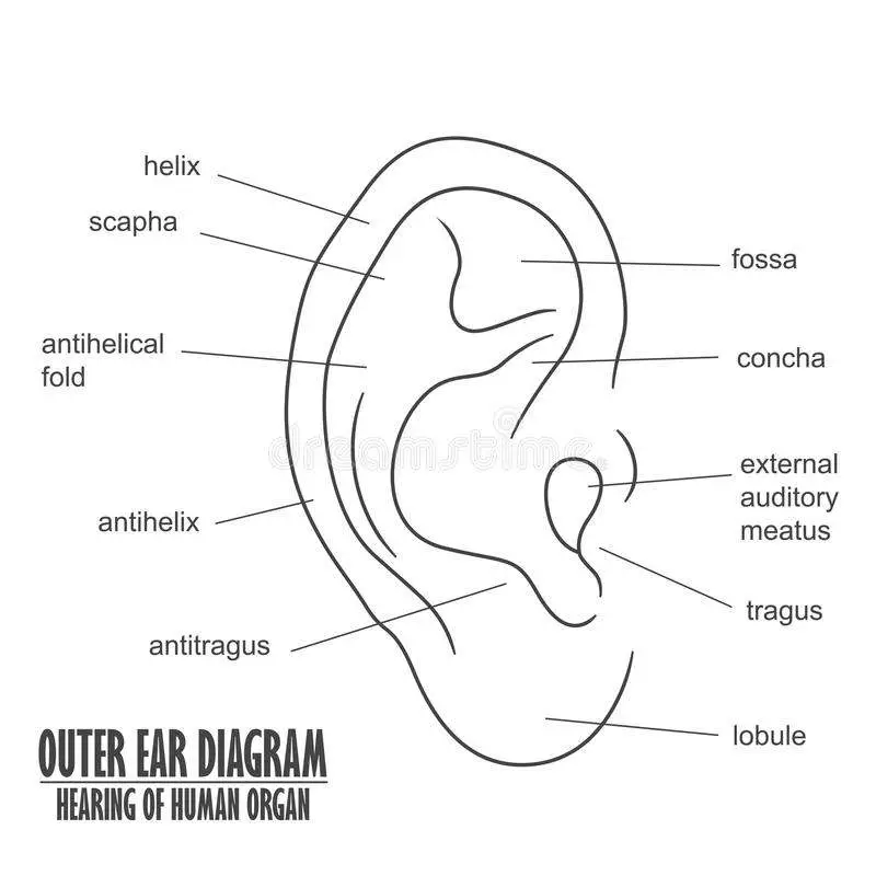
Outer ear diagram
Figure 1.Anatomy of the external ear. 4 Innervation of the auricle. The auricle has several sources of sensory innervation:. The superficial surface is supplied by the great auricular nerve and lesser occipital nerve, both of which are branches of the cervical plexus (C2 & C3), and the auriculotemporal branch of the mandibular nerve, which is a branch of the trigeminal nerve (cranial nerve V)
SPEECH LANGUAGE PATHOLOGY & AUDIOLOGY HEARING DISORDERS OF THE OUTER EAR
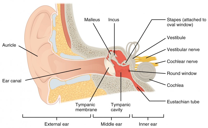
Human ear. The ear is divided into three anatomical regions: the external ear, the middle ear, and the internal ear (Figure 2). The external ear is the visible portion of the ear, and it collects and directs sound waves to the eardrum. The middle ear is a chamber located within the petrous portion of the temporal bone.
Common balance disorders Hearing Link
The diagram of the ear is important from Class 10 and 12 perspectives and is usually asked in the examinations. A brief description of the human ear along with a well-labelled diagram is given below for reference. Well-Labelled Diagram of Ear. The External ear or the outer ear consists of; Pinna/auricle is the outermost section of the ear.
The Ear CP Blas de Otero
I mean the entire structure of the ear. (classical music) So let's look at how the different parts of the ear work together to make us experience sound. So our ear can be divided into three parts: the outer ear, the middle ear, and the inner ear. The outer ear starts with the pinna. It's the part you can see and touch.

Ear Diagram Leaving Cert Human Anatomy
Ear cartilage structures are part of the outer ear, which is also called the external ear in medical and anatomy textbooks. The following ear diagram depicts the inner ear, which contains sensory.

How The Ear Works
The human ear, like that of other mammals, contains sense organs that serve two quite different functions: that of hearing and that of postural equilibrium and coordination of head and eye movements. Anatomically, the ear has three distinguishable parts: the outer, middle, and inner ear.The outer ear consists of the visible portion called the auricle, or pinna, which projects from the side of.
What is conductive hearing loss? Blog of Kiversal
Ear Anatomy - Inner Ear. Ear Anatomy Schematics. Ear Anatomy Images. Chapter 4 - Fluid in the ear. Fluid in the ear Discussion. Fluid in the ear Outline. Middle Ear Ventilation Tubes. Fluid in the ear Images. Chapter 5 - Traveler's Ear.